Eye Examination

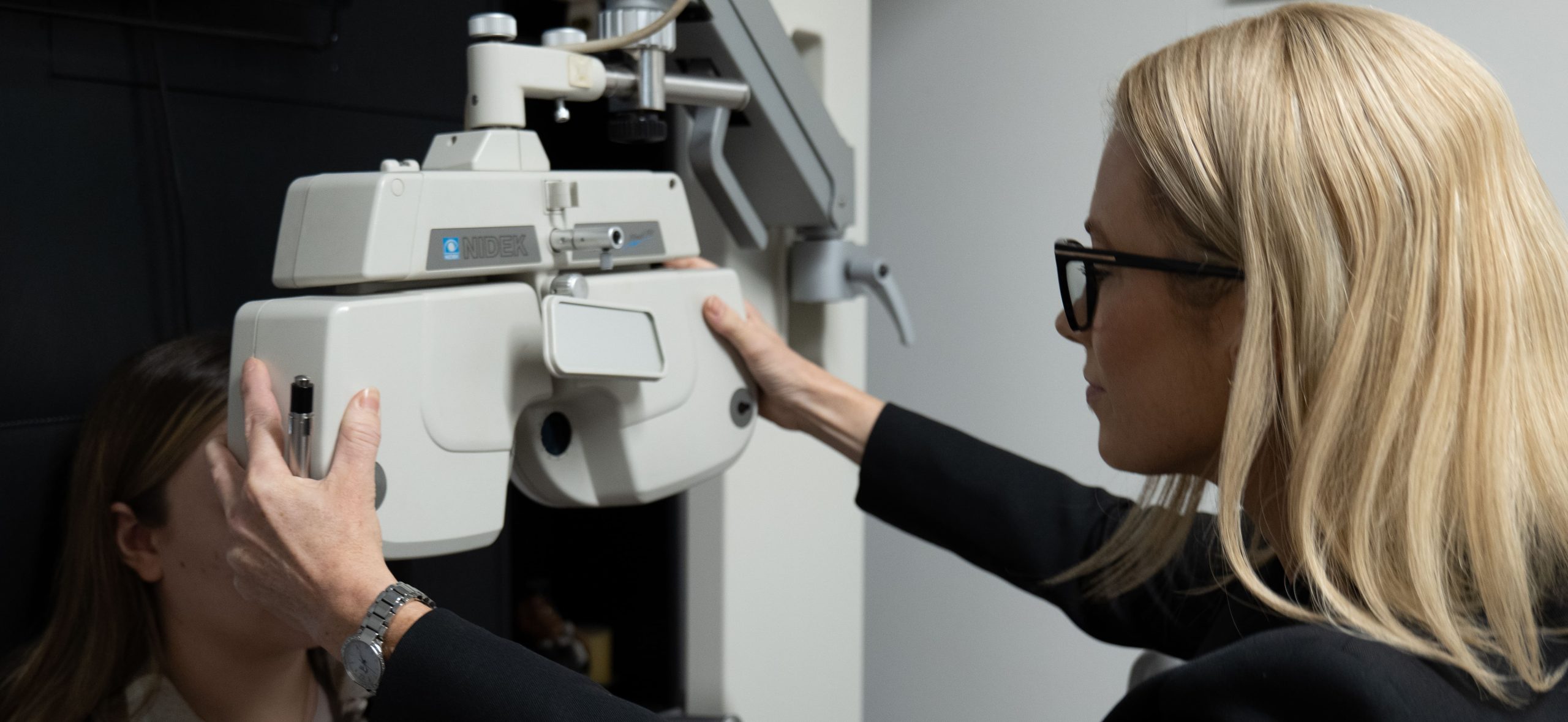
Regular eye examinations are important for maintaining optimal vision, eye health and overall well-being. Booking an eye examination at our optometry practice ensures that you receive personalised care from our experienced optometrists where they not only assess your visual acuity, visual function and determine your prescription, but also screen for potential sight threatening eye conditions. Detecting these issues early can significantly enhance treatment outcomes and prevent further complications. Investing in regular eye examinations is a proactive step towards preserving your eyesight and enjoying a clear and comfortable visual experience. Our recommendation is an eye examination every two years. More frequent exams can be recommended by our optometrists in certain situations where regular observation is required. Trust our optometrists to provide thorough and professional eye care tailored to your individual needs.
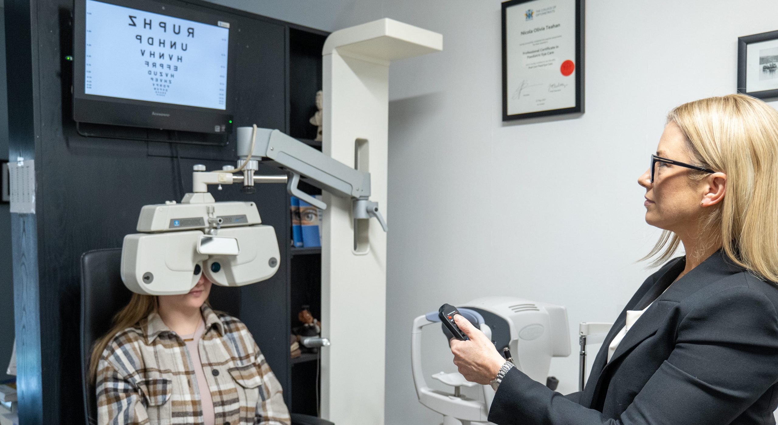
Begin your journey with our expert optometrists who take the time to understand your unique visual needs, preferences, and concerns. Our comprehensive consultations delve into your concerns and lifestyle, offering insights into potential factors affecting your vision, eye health and overall well-being.
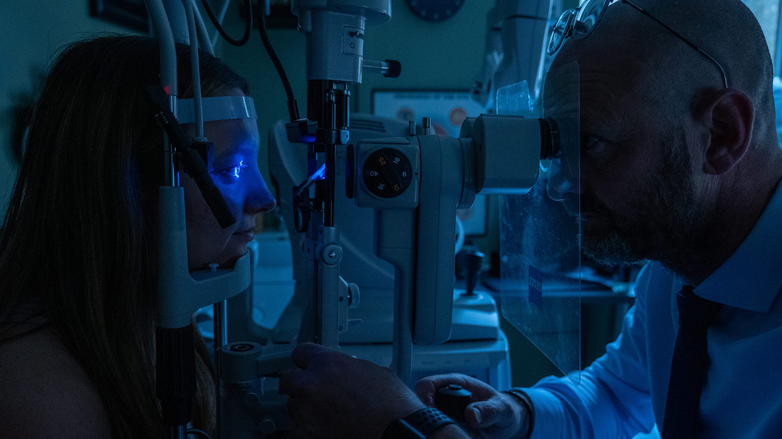
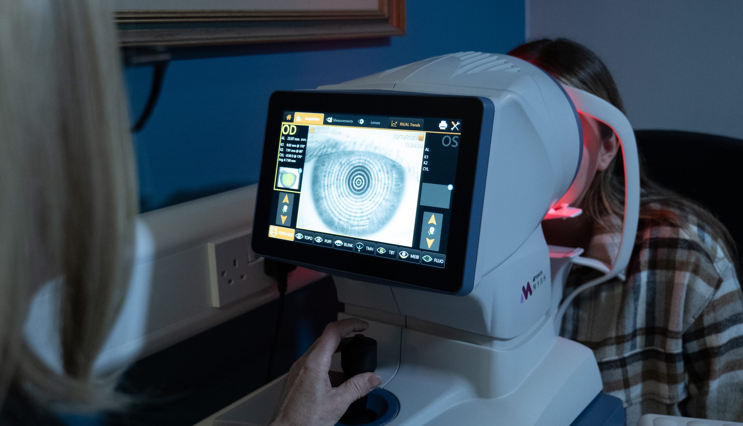
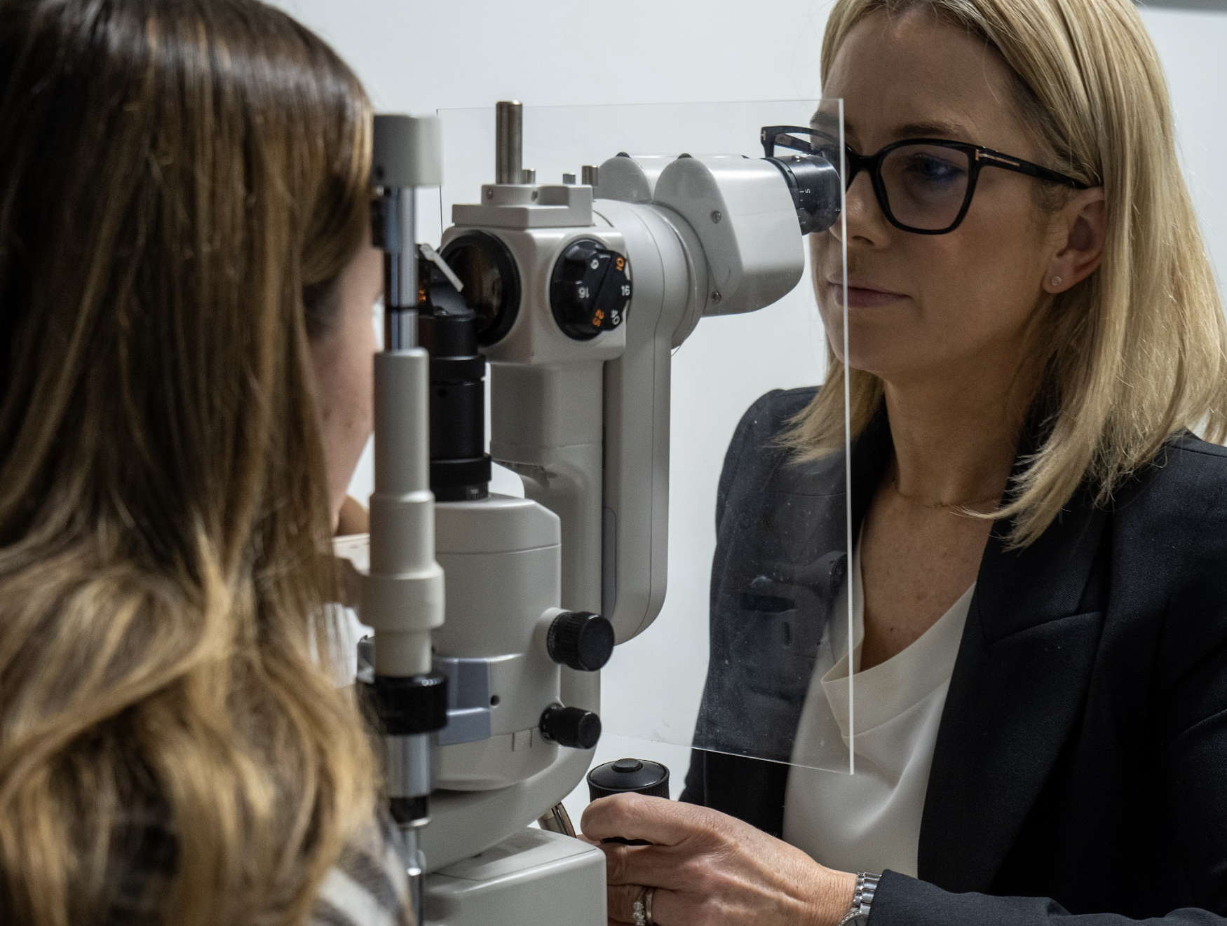
Our optometrists meticulously examine every detail of your eye health, detecting conditions such as cataracts, macular degeneration, retinal disease, glaucoma and more. With us, you can trust that no aspect of your eye health is overlooked.
Did you know that an eye examination can be a window to your overall health? Our optometrists are trained to recognize signs of systemic health conditions such as diabetes and hypertension during your eye examination. Early detection can lead to timely intervention and improved health outcomes.
Any person with symptoms such as blurred vision or double vision, unexplained headaches, eye pain, eye strain, redness, pressure or discomfort in or around the eyes should have an eye examination. Awareness of unusual flashing lights and/or floating spots in the vision should be investigated also.
Often people can require visual reports for job applications, VDU usage at work or driving, and these reports can be provided.
Even if there are no visual symptoms, an eye examination is recommended for anyone who has never had their eyes examined before or has a family member with eye problems. Regularly computer users should have an eye examination every 2 years.

For many people the eye exam can be free of charge. The inclusive scans

  • Medical card holders are eligible for an eye exam every two years. OCT 3D scans are not covered by the medical card allowance and a fee of €40 is applied for this. You may opt out of having the OCT 3D scan. Ask a member of our team how to avail of your entitlements.
  • Self-employed and employees who pay PRSI can avail of an eye exam under the treatment benefit scheme. OCT 3D scans are not covered and a fee of €40 is applied for this. You may opt out of having the OCT 3D scan. Ask a member of our team how to avail of your entitlements.
  • Private health insurance often have day-to-day benefits which may cover your eye examination. Check with your provider to see if you can avail of this benefit and send them your receipts to claim.
Vision is tested using computerised randomised letter charts and automated lens devices called phoropters.

Eye health is assessed using a slit-lamp biomicroscope, a contact tonometer to measure eye pressure, and OCT 3D imaging. The slit-lamp provides a highly magnified view of all anterior structures of the eyeball, including the tear-film, cornea, anterior chamber, iris and crystalline lens. Lids and lashes and the surrounds are also examined. With an additional Volk lens, the examiner can have a 3D natural view to the back interior of the eye which includes the vitreous, macula, retina and optic nerve head. With the OCT 3D imaging, images of the macula, retina and optic nerve head can be captured in 3D and computer analysed for abnormalities.

Eye pressure is measured as a part of the eye health assessment, using an iCare tonometer or Goldmann applanation tonometry. The iCare tonometer is a device that quickly and easily reads the fluid pressure of the eye with minimal sensation.

Keratoconus and corneal topography

Keratoconus is a condition whereby the cornea (the front surface of the eye) thins and warps into a “cone” shape leading to visual distortion. Using a specialised corneal topographer we can map out and monitor the progression of this eye dystrophy and other similar dystrophies, and use the parameters it provides to design a specialised contact lens that will help to minimise the visual distortions the patient perceives. The topographer also aids with the design of our ‘Night’ lenses which are lenses designed to be worn overnight to create a flattening effect of the cornea, giving clear unaided eyesight by morning. Contact our team to learn more about these amazing ‘Night’ lenses.

Anterior eye photography

Any unusual features of the front surface of the eye can be captured and stored using this technique. It is a vital tool in monitoring progression of certain eye problems including cataracts and corneal degenerations. 

OCT 3D scans of retina

Optical coherence tomography (OCT) is a non-invasive, non-contact, transpupillary imaging technique able to produce high-resolution images detailing the 3D structure of the eye, from the anterior segment to the posterior pole. OCT uses light waves to take cross-section pictures of the retina, the very important layer at the back inner lining of the eye. With OCT, your optometrist can view all of the retina’s distinctive layers and they can map and measure the thickness of the retinal layers. Incredible detail of the retinal vasculature can be observed with the digital camera setting. Eye abnormalities such as retinal tears, macular holes and diseases including age-related macular degeneration  and diabetic eye disease can be easily detected using this technology.

Our OCT 3D imaging device can also measure corneal thickness and dimensions, and allow us to view the anterior irido-corneal angle, which is useful in assessing for closed angle glaucoma.

Set your categories menu in Theme Settings -> Header -> Menu -> Mobile menu (categories)
Create your first navigation menu here
Shopping cart
Start typing to see posts you are looking for.