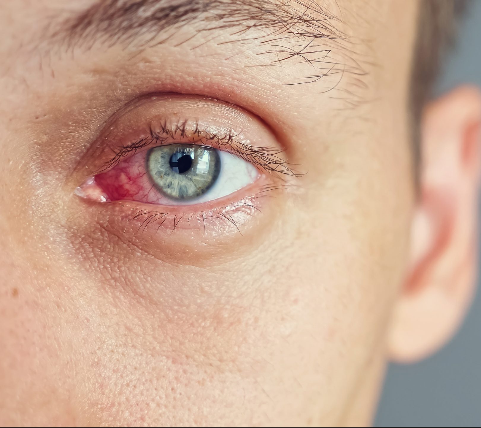
Minor Eye Emergencies
Providing urgent care for minor eye emergencies
At our optometry practice, we understand that unexpected eye issues can arise, demanding prompt attention. For instances of sudden onset problems affecting one or both eyes, we offer immediate assessments without the need for a comprehensive eye examination. Whether it’s a sudden blur, red eye, sore eye, or the discomfort caused by a foreign body like lash, dust, grit, or garden material, our team is equipped to assess and manage these cases efficiently. Rest assured, we can often address these concerns without the necessity of a visit to the A&E eye hospital. And if a visit to the A&E is deemed appropriate, we can send the relevant referral information through our direct referral pathways to the emergency ophthalmology teams, thereby ensuring swift emergency care for those that need it.


